MICROSURGEON.ORG

atlas of microsurgery techniques and principles

Extensor Digitorum Brevis Brevis Flap

Anatomic considerations
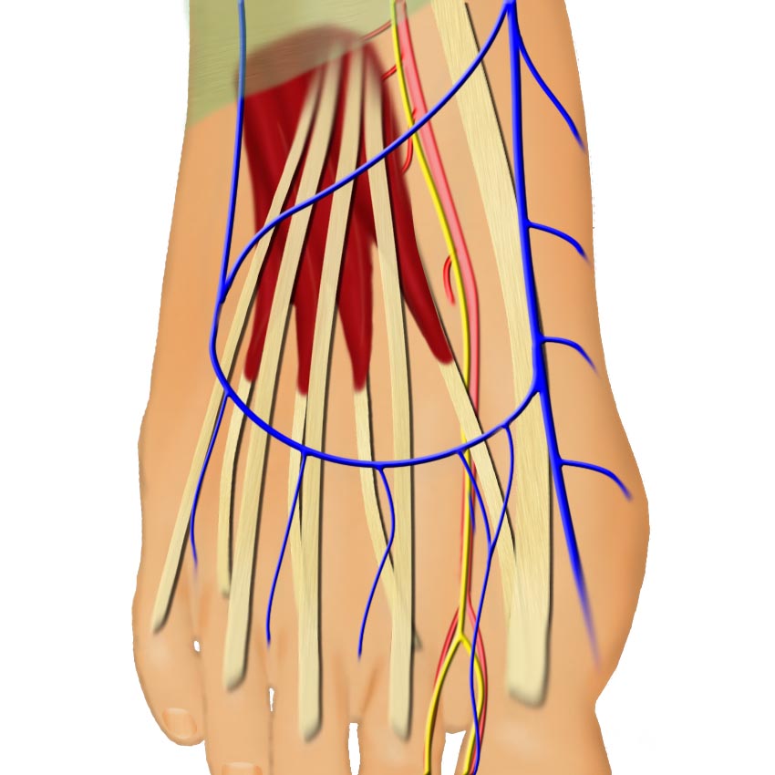
Innervation:
A muscle flap approximately 4 by 5 or 6 centimeters. It is not usually taken with a skin paddle since the muscle lies deep to the long extensor tendons of the foot.
Origin: Anterior surface of calcaneus and inferior extensor retinaculum.
Insertion: The long toe extensors and the proximal phalages of the toes.
Innervation:
Yes - deep peroneal nerve.
Blood supply:
Lateral tarsal artery and direct branch to extensor hallucis brevis from the dorsalis pedis.
Artery:
Large - can be traced to the dorsalis pedis and anterior tibial artery.
Veins: The venae of the dorsalis pedis or anterior tibial artery.
Pedicle length:
Can be made very long by tracing proximally on the anterior tibial system.

Anatomy

The extensor brevis muscle has proximal muscle bulk and 4 tendinous insertions into the long toe extensors. The body of this muscle originates on the talo-calcaneal ligament and the inferior calcaneus on the lateral foot. The extensor hallucis brevis (the most medial slip) inserts on the proximal phalanx of the great toe. The extensor digitorum brevis (the lateral three slips) inserts on the second, third, and fourth toe extensor tendons. This muscle lies just deep to the longus extensor tendons.

Extensor brevis anatomy

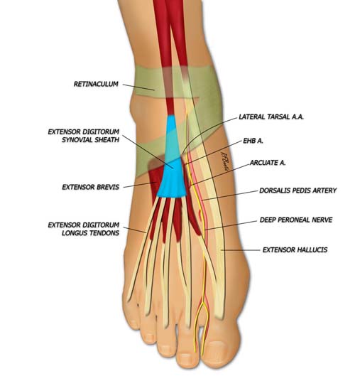
Two lateral branches from the dorsalis pedis artery supply the extensor brevis muscle: the lateral tarsal artery and the artery to extensor hallucis brevis, respectively. The muscle is innervated by a branch of the deep peroneal nerve.

*You have to log in to view the rest of this page. Registration is free.