Flap and Replant Perfusion Monitoring
Monitoring circulation of flaps and replants post-operatively is critical to success in microvascular surgery. To be effective, changes in perfusion need to be recognized quickly to correct any treatable problems. Disruption of perfusion to a flap or replant can result in partial or complete tissue loss. Failure to establish re-perfusion of blood supply after blood vessel repair to an ischemic organ is known as the no-reflow phenomenon. The mechanism is believed to be related to blood vessel injury, specifically endothelial injury, as well as platelet aggregation and leakage of intravascular fluid. The severity of this effect is correlated with ischemia time - the total time the flap lacks perfusion and oxygen flow. Monitoring and quick recognition of ischemia reduces the chance of a no-reflow phenomenon and flap failure.
Clinical Evaluation
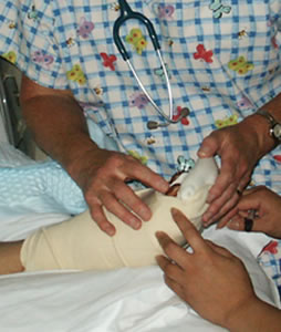 Clinical evaluation by an experience microsurgeon is considered the gold standard for perfusion assessment. Clinical evaluation requires no specialized equipment, but does rely heavily on the experience of the evaluator. Important physical signs include the quality of capillary filling, bleeding from a cut edge of replanted or transferred tissue, and tissue turgor. A pale, cool digit without capillary refill implies an arterial inflow problem. A rigid, blue digit with rapid refill implies venous insufficiency. A pink color with 1-to-2-second refill is consistent with good perfusion. If the nail bed has been exposed, or if a wound edge or exposed tissue surface is accessible, abrasion should cause bright red bleeding. If no bleeding is noted, arterial occlusion is implied. If brisk, dark bleeding occurs, venous insufficiency should be considered. Stabbing with a needle or scalpel yields similar information, but if done repeatedly may cause significant tissue trauma. Turgor in well-perfused replanted digits should approximate the turgor of normal digits. Flaccidity indicates arterial occlusion, while swelling suggests venous obstruction
Clinical monitoring is effective, but can be difficult, and imperfect, even with the most experienced observer. Often it can be tricky to tell if a flap has a circulatory problem. The examiner looks at flap color and sometimes capillary refill or temperature depending on the setting. In addition, there are confounding factors that interfere with assessment by even the most experienced clinician. For instance, replanted fingers are stained with dark colored grease embedded into the skin folds and fingerprints before an accident amputated fingers. Patients with low hemoglobin levels are pale. Flaps and replants are often bled or medicinally leeched to alleviate or attempt to pre-empt venous obstruction, leaving the skin stained with blood and difficult to assess. These and other factors make clinical evaluation often unsatisfying despite being the accepted gold standard in detecting replant and flap compromise.
Flap circulatory disturbances can be divided into arterial insufficiency and venous insufficiency. If there is an arterial circulatory problem the flap would usually look pale and lack capillary refill. Muscle flaps can be particularly difficult to judge – color change with loss of a beefy red appearance is most common. If venous clot is the cause of flap failure, the flap generally becomes congested and bluish in color. Capillary refill is brisk. Sometimes poking a flap with an 18 gauge needle (away from the pedicle site) can help you judge flap circulation. If there is no bleeding, the problem is inflow. If there is rapid exit of dark red blood, venous congestion is likely the problem.
More sophisticated devices to monitor flap flow have been developed. The ideal monitor is reliable, accurate, simple to operative, continuous and inexpensive. Several different monitoring devices are in use now, and include:
- Surface or pencil Doppler
- Temperature probe
- Laser Doppler probe
- Quantitative fluorometer
- Implantable Doppler
- Near infra-red spectroscopy
- Qualitative indocyanine green
Surface or pencil Doppler
 The pencil Doppler probe operates a with a small hand held probe attached to a control box and speaker. The probe is applied to the skin overlying the blood vessel and the presence or absence of a vascular signal is determined by sound. The device has significant operator dependence, although the skill and experience to obtain a signal are not great, interpretation relies on clinical acumen. Not all flaps and replanted parts have Doppler signals that can be obtained, limiting the device to use in a subset of patients.
Quantitative fluorometry
 Quantitative fluorometry is used to monitor replanted parts and tissue transfers with cutaneous components or skin islands. The readings are obtained by nurses. Baseline skin readings are obtained from the monitored tissue and a control area. Repeated readings are obtained 10 and 60 minutes after injection of 0.5 to 1.0 mg/kg of fluorescein intravenously. Absence of a rise in fluorescence at 10 minutes indicates arterial occlusion. Absence of a fall in fluorescence at 60 minutes indicates venous obstruction. Determinations are made every 2 hours in the early postoperative period. Fluorometry often detects vascular complications before physical examination.
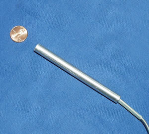
Implantable Doppler
![]()
The implantable Doppler has been found to be highly reliable with several of the qualities needed for an ideal monitoring system. In a study performed by Kind, Buntic and Buncke et. al. in 1998, the implantable Doppler probe was able to salvage 100% of flaps that were failing due to clotting at an anastomotic site. The probe is placed on the draining vein of the free flap to monitor venous flow post-operatively. If flow loss is detected, it may represent venous or arterial clotting in the micro-circulation of the flap and require urgent exploration. Drawbacks include possible tethering of the probe wire causeing venous kinking, difficulty/inability to use in small veins and the qualitative nature of interpreting the signal.
Pulse oximetry
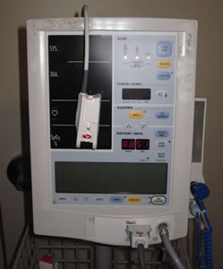 Pulse oximetry detects differences between wavelengths of light absorbed by reduced and oxygenated hemoglobin. The device also records the pulsations of the vascular bed. Pulse oximetry thus continuously monitors both oxygen saturation and pulse. This system, is used widely in hospitals, is reliable, simple to apply, and does not require a sophisticated observer. If a pulse is absent as in an arterial occlusion, or oxygen saturation is decreased as in venous occlusion, an alarm sounds. The monitored tissue, however, must be of the correct shape to allow placement of the probe, which was designed to be placed on digits. Data from the one of the busiest microsugical units in the world have shown this instrument does not promptly detect subtotal venous occlusion, and that the probe adhesion and sensitivity can be compromised by bloody surfaces. It cannot be used in flaps and the probe itself obstructs the clinical assessment of replanted parts.
Near infra red spectroscopy
 Like pulse oximetry, near infra red spectroscopy detects the reflected spectra of oxygenated and deoxygenated hemoglobin in the microcirculation of the skin. The skin is illuminated by the photometer's fiberoptic light and the reflected light is analyzed by a photodiode. Oxygenated hemoglobin has two peaks in its extinction spectra. With deoxygenation, the peaks decrease in amplitude and begin to merge. A microprocessor calculates oxygen saturation based on the difference in the distance between the peaks. In practice the device numbers widely vary and are difficult to correlate clinically. The probe and fiber optic attachment are quite stiff and obstruct replanted parts if used continuously.
Temperature monitoring:
 Cutaneous temperature monitoring measures skin temperature as an indicator of perfusion of the monitored tissue and has been used in many experimental and clinical settings. With adequate perfusion, the monitored tissue surface temperature should be constant and within a few degrees of a well perfused control area. Changes in core temperature and in ambient temperature and heat retention by dressings cause spurious readings, negating the premise of the technique.
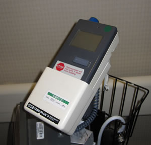
Arteriography
 Arteriography can demonstrate the patency of vascular anastomoses, but is limited as a postoperative monitoring technique because it cannot be done frequently enough to be a practical routine. It is invasive, requires a large team and exposes the patient to contrast dyes. Its use in the very small blood vessels of microsurgical patients is difficult and visualization of these small vessels is poor.
Indocyanine green
 Indocyanine green has been used to detect vascular perfusion in coronary vasculature with great success. However, its role in postoperative monitoring of flaps and replants is still to be determined. Currently available technology is large, lacking ease of portability for ward assessment, and requires skilled physician interpretation, like arteriography. And like arteriography, it is llimited in the postoperative setting since it cannot be done frequently enough to make it routine.
Laser Doppler
Laser Doppler measures capillary blood flow by calculating changes in the wave length of laser light reflected off moving red cells. In measuring tissue perfusion, the laser Doppler registers the presence or absence of red-cell motion in dermal capillaries and cannot distinguish between arterial and venous occlusion. Surface probes are fixed to the skin and provide continuous monitoring. Problems with probe applications include tissue compression caused by adhesive strips or loss of probe contact on moist or bloody surfaces. False-positive readings are frequent and the use of Laser light is a drawback. Use on replanted parts is limited by the small size of the parts relative to the probe and requirement for the probe to be fixed to the parts, obstructing the clinical assessment of the part.