Helical Rim Flap
Anatomic considerations
Authored by Thomas E. J. Hayakawa, MD, FRCS(C)
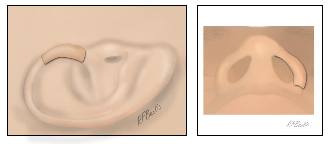
Full thickness loss of the nasal ala is difficult to reconstruct. As pointed out by Pribaz in 1993, the ascending helical rim has an uncanny resemblance to the nasal ala. It also provides skin and cartilage, as well as a comparable color match to provide for reconstruction of this very prominent area, while leaving an aesthetically pleasing donor area.
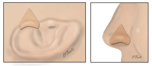
The anatomy of the ascending helical rim and nasal ala have an uncanny similarity (Pribaz and Falco, Ann Plast Surg, 31:289, 1993).
The similarity in the helical rim and nasal ala from the inferior nasal view.
Vascular Anatomy
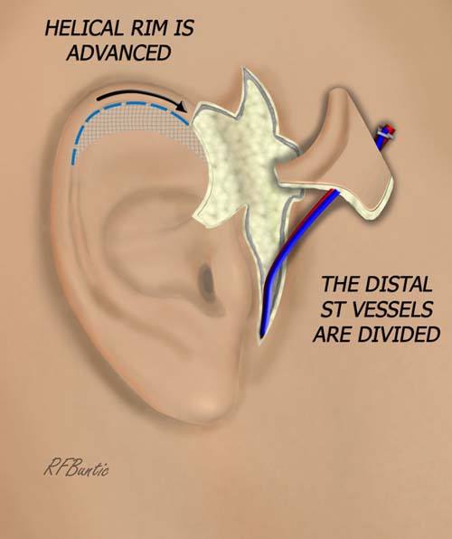
The superficial temporal vessels ascend superiorly, just anterior to the ear as the travel toward the scalp and superficial temporal fascia. During this course, the vessels distribute numerous small branches including branches to the ascending helical rim. The superficial temporal vessels are the basis of this flap, with the soft tissue resection capturing inflow and drainage to this flap via small branching vessels.
The anatomy of the superficial temporal vessels can be aberrant on occasion, particularly the vein. We have found occasion where the superficial temporal vein is not present in proximity to the artery.