Medial Femoral Condyle Flap
The Anatomic considerations
The medial femoral condyle flap has been noted by Shin to be an excellent source of vascularized bone for treatment of complex bone defects necessitating vascularized osseus reconstruction. The anatomy is consistent, straight forward and provides a large block of corticocancellous bone well suited to reconstruction of defects in both the upper and lower extremities as well as the mandible.
Anatomy
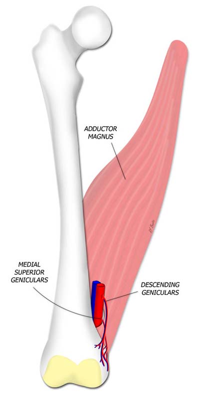
Arterial inflow to the medial femoral condyle flap is supplied by the descending genicular artery, a branch of the superficial femoral artery. This branch runs deep in the medial thigh, deep to the vastus medialis, and coalesces with the medial superior genicular artery to supply a vascular plexus to the medial femoral condyle.
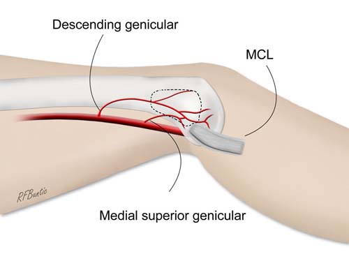
The medial femoral condyle is supplied by a plexus of vessels from the descending genicular artery and the medial superior genicular artery. The flap is marked by identifying the vascular plexus on the medial condyle and incorporating a component of the network in the flap. The medial collateral ligament (MCL) and joint capsule of the knee are spared and not incorporated into the flap.