Extensor Digitorum Brevis Brevis Flap
Anatomic considerations
Anatomy
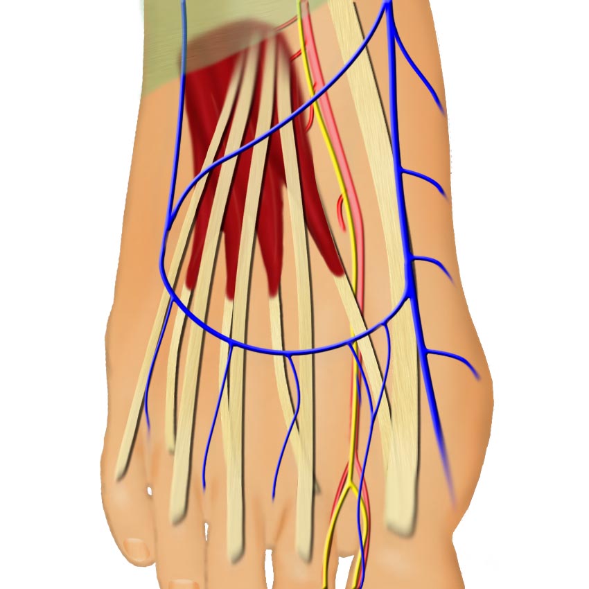
The extensor brevis muscle has proximal muscle bulk and 4 tendinous insertions into the long toe extensors. The body of this muscle originates on the talo-calcaneal ligament and the inferior calcaneus on the lateral foot. The extensor hallucis brevis (the most medial slip) inserts on the proximal phalanx of the great toe. The extensor digitorum brevis (the lateral three slips) inserts on the second, third, and fourth toe extensor tendons. This muscle lies just deep to the longus extensor tendons.
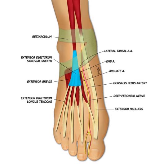
Two lateral branches from the dorsalis pedis artery supply the extensor brevis muscle: the lateral tarsal artery and the artery to extensor hallucis brevis, respectively. The muscle is innervated by a branch of the deep peroneal nerve.
The lateral tarsal artery, a branch from the dorsalis pedis artery supplies the extensor hallucis brevis and extensor digitorum brevis muscles. It originates under the extensor retinaculum and enters at the proximal aspect of the muscle belly.
A branch of the deep peroneal nerve innervates the extensor brevis muscle. This branch parallels the course of the lateral tarsal artery and enters at the junction of the proximal and middle thirds of the muscle belly. Although this nerve branch is short, intraneural dissection under microscopic magnification allows additional length.
Flap Harvest

A dorsal incision is made over the course of the anterior tibial artery and extended in a soft curve over the muscle..
Harvest of the muscle is performed with the patient in the supine position with an optional thigh tourniquet. The course of the dorsalis pedis artery should be identified by Doppler probe. Palpation before the induction of anesthesia while the patient extends the toes identifies the body of the muscle over the foot. A lazy-"S" incision is made on the axis of or just lateral to the anterior tibialis pulse at the ankle and extends onto the dorusm of the foot.

The long extensors are identifed and retracted laterally. The dorsalis pedis artery is traced under the divided estensor retinaculum and the arterial branches to the extensor brevis are identifed.
Branches of the superficial saphenous system are ligated as necessary. The skin flaps are developed in a plane just above the peri-tenon of the long extensor tendons.

The muscle is elevated from medial-distal to proximal-lateral.
The long extensor tendons are retracted exposing the short extensors and muscle bellies. The extensor hallucis brevis tendon is then elevated and retracted from where it crosses the first metatarsal space exposing the dorsalis pedis artery, venae, and deep peroneal nerve. The extensor brevis tendon insertions are identified and transected. They are then controlled as a group so that individual muscle bellies are not avulsed from each other. The flap is then elevated from distal to proximal.
The nerve to the flap can be taken with the deep peroneal nerve, or is transected before flap insertion. The dorsalis pedis vessels are ligated and divided before the takeoff of vessels to the brevis muscles. As the dissection progresses the muscle is turned over idenfifying the entry of the pedicle under the muscle and the origin of the muscle. The muscle origin is freed sharply or with a fine cautery on low setting. The necessary artery and vein length is gained by tracing the dorsalis pedis system to the anterior tibial vessels.
After hemostasis is obtained, the donor site is closed over a suction drain .
Post-Operative Care
The leg is strictly elevated for two weeks. After that point, compression with a compression bandage is employed whenever the leg is dependent for an additional two weeks.