Medial Femoral Condyle Flap
The Anatomic considerations
The medial femoral condyle flap has been noted by Shin to be an excellent source of vascularized bone for treatment of complex bone defects necessitating vascularized osseus reconstruction. The anatomy is consistent, straight forward and provides a large block of corticocancellous bone well suited to reconstruction of defects in both the upper and lower extremities as well as the mandible.
Anatomy
Arterial inflow to the medial femoral condyle flap is supplied by the descending genicular artery, a branch of the superficial femoral artery. This branch runs deep in the medial thigh, deep to the vastus medialis, and coalesces with the medial superior genicular artery to supply a vascular plexus to the medial femoral condyle.
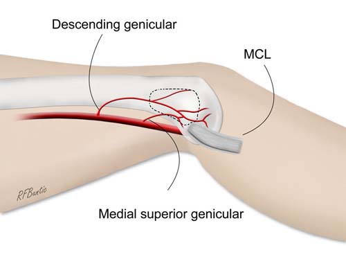
The medial femoral condyle is supplied by a plexus of vessels from the descending genicular artery and the medial superior genicular artery. The flap is marked by identifying the vascular plexus on the medial condyle and incorporating a component of the network in the flap. The medial collateral ligament (MCL) and joint capsule of the knee are spared and not incorporated into the flap.
The medial superior genicular is a shorter branch, and although the flap could be taken on this branch, it is preferably taken on the descending genicular to supply a longer vascular leash.
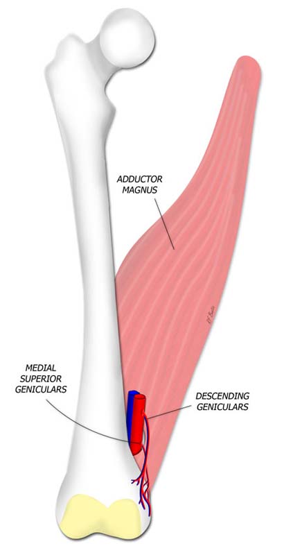
The femoral artery and vein pass through the hiatus in the adductor magnus to enter the popliteal space and become the popliteal artery and vein respectively. The descending geniculars arise from the femoral system before the popliteal space, while the medial superior geniculars arise near the level of the muscle hiatus. The adductor magnus is on the floor of the exposure of the geniculate vessels and the medial condyle of the femur.

Exposure of the flap requires identifying the vastus medialis muscle and retracting it anteriorly to expose the medial femur and medial femoral condyle. The sartorius, gracilis and hamstrings are all retracted posteriorly. On the floor of the dissection is the femur, femoral vessels and the adductor magnus muscles.
Flap Dissection
The patient is supine in the frog legged position or in the lateral decubitus position. A tourniquet is optional but helpful. The dissection can be quite deep and the pooling of blood in the deep wound can be a nuisance to dissection if no tourniquet is present.
A medial thigh incision is made extending from the medial femoral condyle proximally along the posterior axis of the femur. During subcutaneous dissection the saphenous vein is identified and retracted posteriorly. The muscular fascia is incised and the vastus medialis is teased from its septal fascial attachments and retracted anteriorly.

An incision is made on the posterior axis of the femur. The saphenous vein travels through the area of the incision and is retracted posteriorly. The muscular fascia is incised longitudinally and the vastus medialis muscle is retracted anteriorly.
On the deep surface of the femur the descending genicular artery is identified and traced distally to the level of the medial condyle. There are several branches arising from the descending genicular vessels and these are ligated and divided.

In the plane between the vastus medialis and the gracilis muscle, the surface of the femur is identified and the genicular vessels are identified. The adductor magnus lies on the floor of the dissection along with the femur and femoral vessels.
The pedicle is freed of deep attachments from its distal portion just proximal to the medial condyle. The area for the bone flap is identified and marked. A rectangle up to 2.5 x 1.5-cm can be harvested.
The perichondrium is incised with the Bovie electrocautery on a low cut level. The cortex of the bone is incised using a micro oscillating saw. Osteotomes are used to elevate the bone flap from the entire perimeter of the corticotomy. Using a curved osteotome, the flap can be removed with some cancellous bone approximately 1-1.5-cm thick.

After corticotomies are made, the flap is elevated with an osteotome using care not to fracture the flap.
The entire flap is then isolated on the pedicle and cancellous bone from the donor defect can be scooped out for any additional non-vascularized bone grafting. The donor area is packed with hydroxyapatite paste and the flap is ligated and divided when the recipient area is ready.
The wound is closed with interrupted deep sutures and skin sutures or staples over a suction drain.

The pedicle can be traced to the origin to gain length.
Post-Operative Care
The patient is allowed to ambulate as soon as clinically indicated for the flap reconstruction. The drain is usually pulled before discharge.
Bibliography
- Kakar S, Duymaz A, Steinmann S, Shin AY, Moran SL. Vascularized medial femoral condyle corticoperiosteal flaps for the treatment of recalcitrant humeral nonunions. Microsurgery. 2011 Feb;31(2):85-92.
- Jones DB Jr, Shin AY. Medial femoral condyle vascularized bone grafts for scaphoid nonunions.
Chir Main. 2010 Dec;29 Suppl 1:S93-103. - Bakri K, Shin AY, Moran SL. The Vascularized Medial Femoral Corticoperiosteal Flap for Reconstruction of Bony Defects within the Upper and Lower Extremities. Semin Plast Surg. 2008 Aug;22(3):228-33.
- Choudry UH, Bakri K, Moran SL, Karacor Z, Shin AY. The vascularized medial femoral condyle periosteal bone flap for the treatment of recalcitrant bony nonunions. Ann Plast Surg. 2008 Feb;60(2):174-80.