Microvascular Lymph Node Transfer
Anatomic considerations
Anatomy
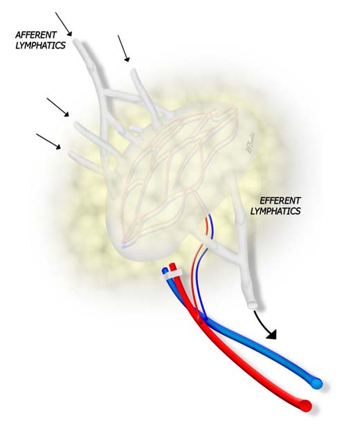
Vascular and lymphatic flow anatomy of a lymph node. The node has both afferent and efferent lymphatics and a vascular pedicle.
Each lymph node has afferent lymphatic vessels that transport lymph to the node for immunological processing, and an efferent system that transports fluid toward the thorax and drainage back into the chylothoracic duct. The node also has an arterial vascular inflow and outflow, and the tissue of the node itself is very well vascularized. Because the node vessels are extremely small, a larger artery and vein must be captured for inflow and outflow in order to perform microvascular anastomosis and transplant the node as a vascularized system. If the node is sufficiently large, then an efferent lymphatic duct may be captured and coapted to a proximal vein to allow efferent drainage directly into the venous system.
Operative Procedure
There is an abundant supply of nodes in the groins bilaterally, and a small sampling of one to three nodes can be removed with a vascular leash for micro lymphatic reconstruction. Most commonly, lymphatic reconstruction is done in the axilla for treatment of refractory upper extremity lymphedema after mastectomy and lymph node dissection.
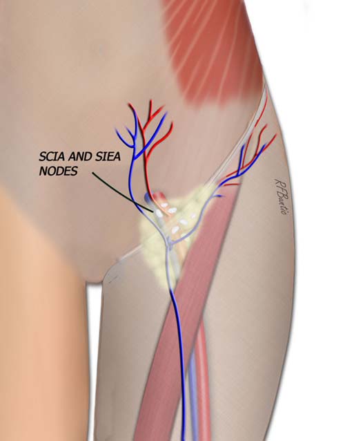
The SCIA or SIEA system can provide a vascular pedicle for a groin node system.
The groin nodes are approached directly over the superficial circumflex or SIEA vessels. Our preference is for the SIEA system since it can be approached through a low transverse abdominal incision.
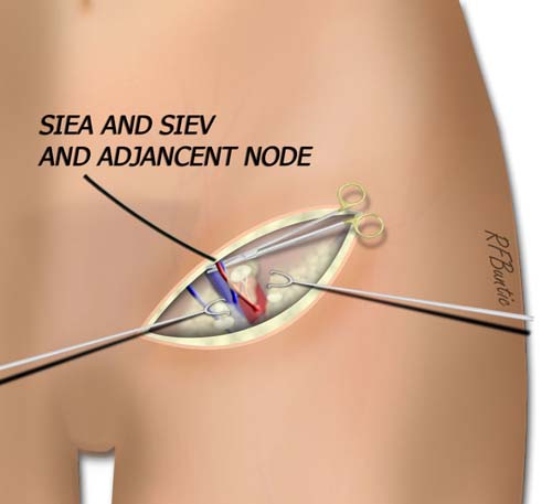
The SIEA vessels are depicted and isolated with adjacent perivascular soft tissue including a lymph node.
The SIEA vessels are found, and a segment of fat with the vessels containing at least one large node is captured. The distal vessel stump can be divided and the vessels, lymph node(s) surrounding fat are isolated on the pedicle.
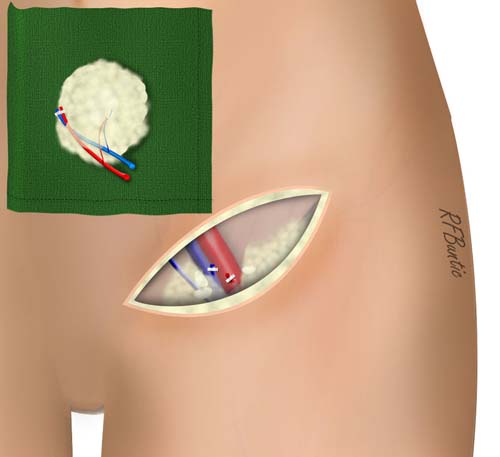
When the flap is isolated, the vascular pedicle can be divided according to the timing needed for the recipient area.