The Serratus Muscle Flap
Anatomic considerations
The serratus anterior muscle takes origin from the lateral scapula and fans anteriorly as it inserts into the first nine ribs. Only a portion of the muscle is used for flap reconstruction: the lower three slips of muscle arise at the very inferior scapula and have an independent blood and nerve supply, making them a good source of a medium to small sized muscle for free flap coverage. One, two or three slips can be harvest, depending on requirements, and the muscle can be neurotized to form a functional unit.
Vascular Anatomy
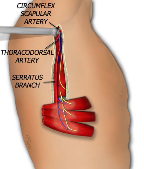
The vascular anatomy of the serratus is related to the latissimus and axillary vessels, familiar territory for the reconstructive surgeon. The subscapular artery, a branch of the third part of the axillary artery, descends inferiorly to supply the serratus artery. Before branching into the serratus it gives off a circumflex scapular branch and the thoracodorsal artery. The superior slips of the serratus are supplied by the lateral thoracic artery, a branch of the second part of the axillary artery under the pectoralis minor.
The long thoracic nerve supplies the lower three slips of serratus, running inferiorly on the surface of the muscle, becoming more adjacent to the vascular pedicle as it descends.
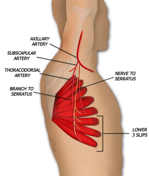
The lower three or four slips of the serratus are used for the flap.

The posterior aspect of the serratus is partially covered by the latissimus muscle.
Operative Procedure
The patient is positioned laterally, similar to the latissimus harvest. The incision is marked extending from near or at the axilla, in a gentle curve anterior to the latissimus that sweeps over the 7th, 8th or 9th rib.
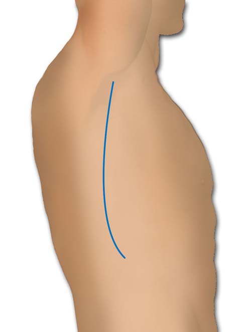
The incision traces the border of the latissimus then curves anteriorly.
The dissection is carried through the muscular fascia, exposing the anterior latissimus and the serratus muscle. The latissimus is retracted posteriorly and the surface of the lower slips of serratus are exposed from their origin on the inferior angle of he scapula to their rib insertions.
The artery and nerve to the serratus are dissected proximally. The nerve to the lower slips is isolated, and proximal extension of this nerve is mobilized by intrafascicular dissection of the nerve to separate from branches to the upper muscle. The nerve supply to the upper slips must be kept intact to avoid winging of the scapula. Depending on pedicle length, the thoracodorsal artery and circumflex scapular artery can be ligated to obtain a long vascular leash.
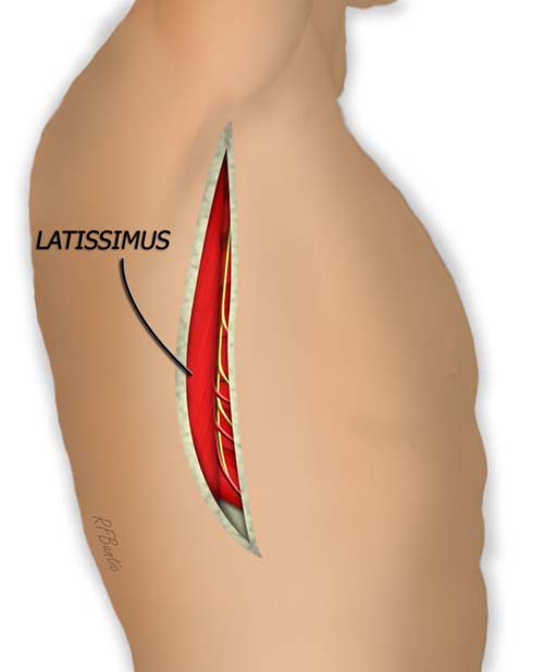
The anterior border of latissimus and lower serratus are identified, and the latissimus can be elevated and retracted posteriorly.
The lower edge of the ninth slip of serratus is identified and an plane is developed below the posterior aspect of the muscle, near the angle of the scapula. The connections between the 6th and 7th slip are separated with the bipolar cautery, while preserving the vascular leash and the nerve supple. The muscle can now be detached from the scapula by placing a finger underneath and transecting the posterior insertion with cautery.
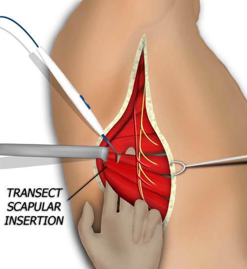
The posterior insertion of the lower 3 slips of the serratus are isolated and divided.
The anterior insertion can then be separated by sharply dissecting the rib insertions of the 7th, 8th and 9th slips. The plane is not distinct and requires cauterizing of small vessels perforating from the intercostals.
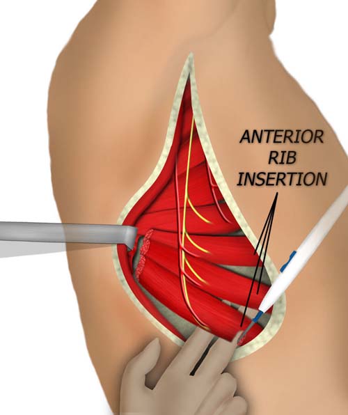
The anterior insertion of the serratus is and also divided. The flap can now be raised from inferior to superior, with all the vascular branches being divided.
The pedicle is isolated up to the subscapular artery if needed for length.
The pedicle can be traced up to the axilla to gain length, if necessary.